
Bringing Augmented Reality to Brain Surgery
Can a piece of paper and a smartphone lead to improvements in brain surgery? That’s the hope of a team based at the Western University in London, ON. Read on to find out more…
Bringing Augmented Reality to Brain Surgery:
Model the structure and function of the brain preoperatively
Neurosurgeons require extensive knowledge of each patient’s brain anatomy, as well as advanced visuo-spatial skills to accurately maneuver surgical instruments during operations. Currently, surgeons look at 2D displays of MRI scans (magnetic resonance imaging) and then mentally transform the patient’s anatomy from the screen to the physical patient (i.e. 3D anatomy). This is particularly challenging in the case of junior trainees who may have limited previous surgical experience and less developed spatial reasoning skills. Ultimately, this leads to longer surgeries and increases the chance of error associated with reduced performance due to the resulting cognitive load.
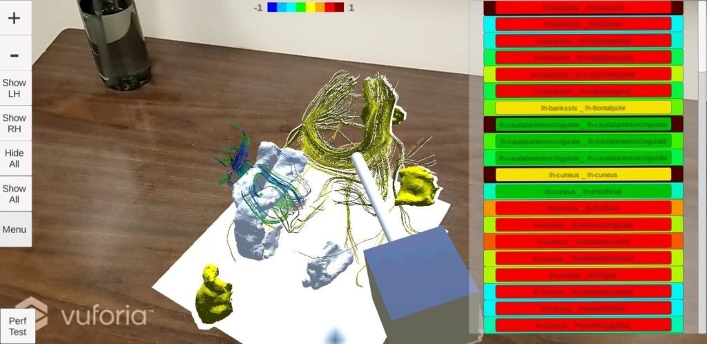
Recently, there has been an increase in the use of Computer-Assisted Interventional approaches, owing in part to advancements in medical imaging techniques. Our research group uses multiple medical imaging techniques (e.g. diffusion weighted imaging, functional magnetic resonance imaging) to gather information about the structure and function of the brain. Using this information, we create 3D models of the brain regions and their structural and functional connections. This approach facilitates surgical trajectory planning. The main challenges associated with current techniques is the lack of intuitive visualization and interactive methods available to neurosurgeons and trainees, due to the fact that they are still rendered on 2D displays.
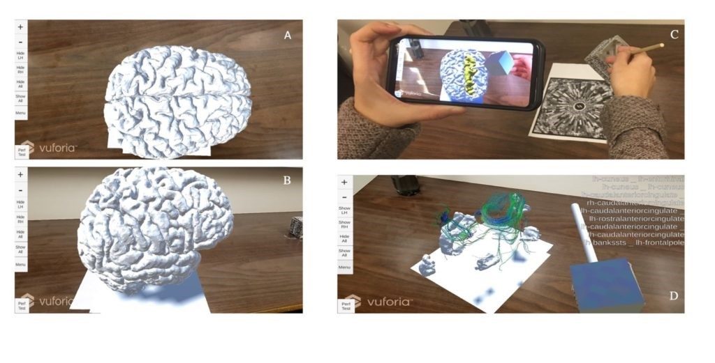
We are developing an augmented reality application which will allow surgeons to blend patient-specific models of the brain derived from preoperative data onto the real world. Augmented reality allows the user to view and interact with digital objects within the real world. Our application is also lightweight requiring only a mobile device and a visual tracking pattern (see picture). The application is designed and evaluated with human factors in mind, and it allows real-time, intuitive, 3D visualization and interaction with brain structures to facilitate training and planning for neurosurgical procedures. We are quantitatively assessing the improvement in targeting performance in terms of the increase in both speed and accuracy. This application allows the neurosurgeon to see the MRIs in 3D and circumvents the 2D to 3D mental transformation for each patient.

Although, our project is still in progress, we have tested it on a sample of students and it is showing promising results. Next, we are looking into different ways to model brain tumours, and eventually have surgeons evaluate the application.
We expect that our application will allow surgeons to safely explore the patient anatomy and identify important areas based on advanced image processing, as well as to plan neurosurgical procedures prior to surgery. We intend to develop our tool as an educational model for neurosurgical resident and medical students for neurosurgical planning.