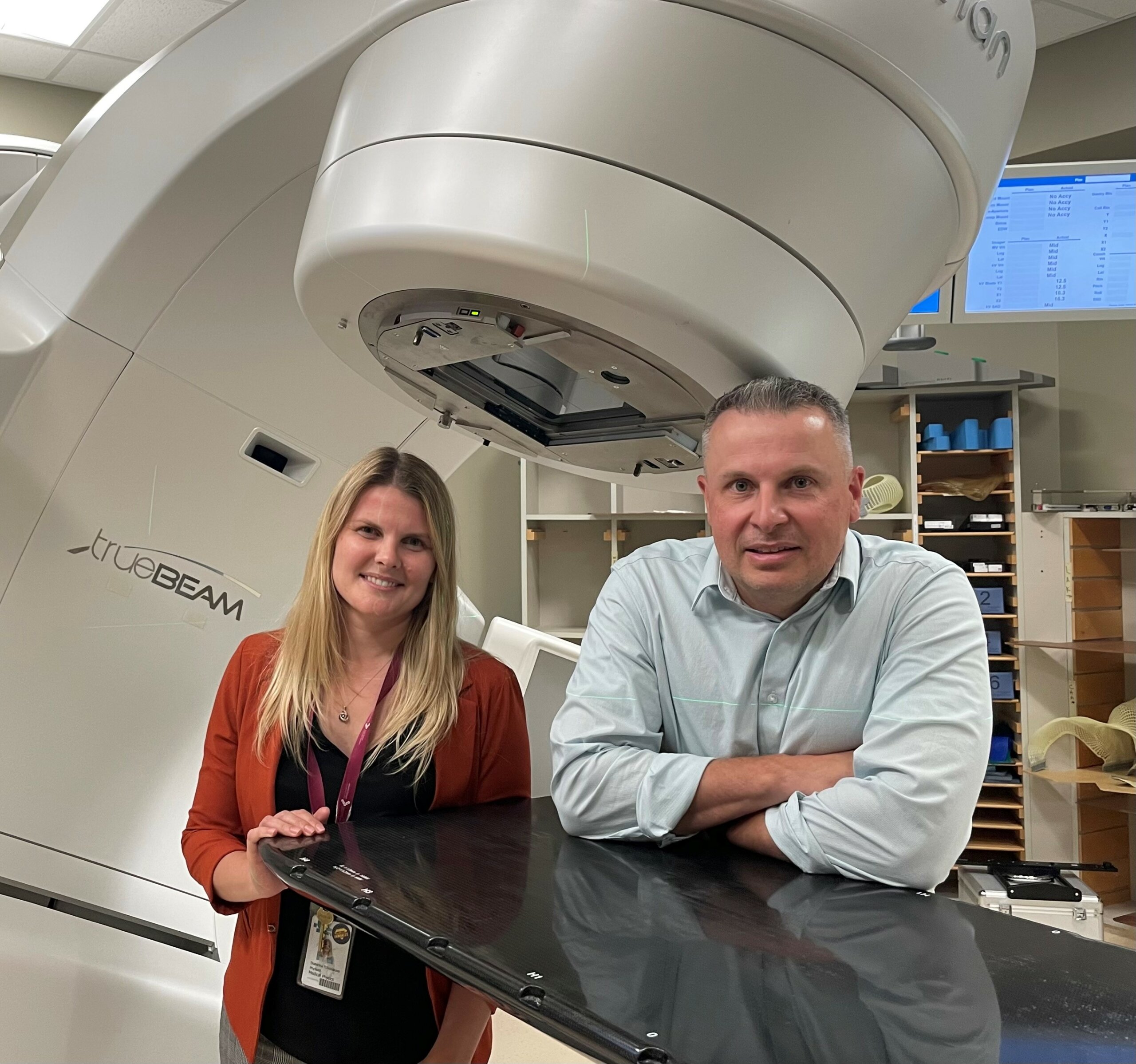
Ekaterina Tchistiakova and Nicolas Ploquin – 2022 Research Grant Recipients
 Ekaterina Tchistiakova and Nicolas Ploquin -University of Calgary, AB
Project Title: “Application of machine learning and diffusion based radiomics for early detection and treatment response assessment of brain metastases”
Description of Project:
Brain metastases are common among cancer patients and lead to significant increase in disease burden. Advanced radiation therapy techniques such as stereotactic radiosurgery can be used to treat brain metastases while sparing healthy tissues and greatly improving patients’ quality of life. Early detection of brain metastases and assessment of treatment response are critical. Currently, radiation oncologists rely on structural Magnetic Resonance Imaging (MRI) for these assessments. Structural changes in the brain take time to manifest on imaging which may delay metastases detection. Other advanced imaging techniques may be more sensitive in detecting microscopic changes prompting closer monitoring of areas at risk, early management and treatment adjustment in case of recurrence. Diffusion weighted imaging (DWI) is an MRI technique which uses patterns of water molecule diffusion to differentiate between healthy and malignant tissues. We will focus on utilizing DWI images from patients undergoing stereotactic radiosurgery treatments. An advanced computing technique called Machine Learning will be used to identify features in DWI allowing for the detection of brain metastases prior to their manifestation on conventional MRI imaging. This methodology will help us develop a framework to achieve the main goal of this project of improving the management of brain metastases in cancer patients.
What receiving this award means:
“Radiosurgery is one of the common treatment options for patients with brain metastases. It has several benefits over other treatment options including reduced side effects, it’s non-invasive nature and improved quality of life especially when administered early. As clinical medical physicists, we are closely involved in the care of cancer patients. We often see the benefits of the new treatment techniques but also where the gaps are and where improvements can be made. One such area is a wider utilization of advanced MRI techniques in radiation therapy. We
are very thankful to Brain Tumor Foundation of Canada for providing us the opportunity to advance our research which investigates the use of DWI MRI and Machine Learning to develop a tool for early detection of brain metastases”.
Midpoint – February 2024
“Brain metastases pose a major challenge in cancer treatment, often leading to diminished patient quality of life and lifespan. Our research is at the forefront of utilizing artificial intelligence to analyze brain images obtained via MRI. We employ diffusion MRI as a specialized tool to examine water movement within the brain, thereby revealing subtle changes in the microstructure of brain tissue during the early stages of cancer development. We’ve successfully published two research papers: the first explores unique patterns in water diffusion, identifying how these can be altered by irregular vasculature and disrupted extracellular space, while the second paper leverages these patterns to feed a machine-learning model that distinguishes between healthy and pre-metastatic tissue. Currently, our efforts are concentrated on refining our machine learning algorithms to pinpoint the specific brain regions most susceptible to future cancer development, with the ultimate aim of transforming our software into a clinical tool for early risk assessment of metastatic spread.”
Final Report – April 2025
Our research focused on the use of brain magnetic resonance images (MRI) from patients who are at risk of cancer spreading, and utilizing artificial intelligence (AI), to detect the spread of cancer early. Since over 40% of cancer patients will develop at least one brain metastasis in their lifetime, detecting these malignant growths earlier would dramatically improve cancer care.
We conducted our research by observing how water molecules behave within the body. In a healthy brain, water flows normally along defined pathways. However, when cancer forms, that flow can be negatively affected and often prevents proper function of the brain. By using a special MRI technique called diffusion-weighted imaging, instead of seeing the overall structure of the brain, we can visualize the movement of water through these structures.
We analyzed diffusion images over time to quantify subtle changes in the flow of water that may occur before brain cancer is visible on the standard MRI. Throughout our work, we found characteristic patterns that tended to occur in areas where cancer began to spread. These complex statistical anomalies were fed into the AI model to learn what metastasis formation looks like. As a result, our models were able to detect potential cancer growth earlier than the current clinical standard.
The resources required for research, such as data collection time and hardware, would not have been possible without the generous support from Brain Tumour Foundation of Canada. It was only through their funding that we were able to develop AI models in a reasonable time frame in-house within the privacy-focused framework of healthcare applications. Our work presents an opportunity to leverage data and recent technological advancements to improve the lives of Canadians battling cancer. By detecting metastases earlier, we aim to give back one of their most precious resources: time.