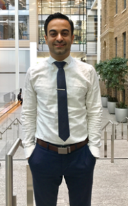
Phedias Diamandis – Research Grant – 2017
Phedias Diamandis, Neuropathologist / Clinician Scientist / Assistant Professor, University Health Network / Princess Margaret Cancer Centre
Project title: “The brAIn Project: biological rendering through Artificial Intelligence and neural networks”
Project Summary
 Brain tumours represent a diverse group of diseases with highly variable therapies and outcomes. A key way to predict how a tumour will behave is by analyzing its specific morphologic features under the microscope. The human eye, however, cannot reliably detect subtle differences. This may explain why patients with the same diagnoses can experience dramatically different outcomes. Although new “molecular” technologies can reliably differentiate between tumour types, molecular testing is oftentimes unavailable and doctors must still rely on microscopic findings to make important clinical decisions.
Recently, advances in “Artificial Intelligence (AI)” allow computers to excel at analyzing images to identify extremely subtle differences. Dr. Diamandis’ team aims to take advantage of AI, and train computers to differentiate between brain tumour types.
Training involves exposing computers to a large series of images with known clinical outcomes (survival, therapy response) to allow AI to “discover” microscopic patterns associated with these specific clinical events. Because computers can process larger amounts of information than humans, it is expected they will be better able to predict tumour behavior from their microscopic appearance. This AI-assisted approach will hopefully allow improved differentiation of brain tumour types and offer more accurate predictors of outcome and treatment response to patients.
Project Update
Brain tumours represent a diverse group of diseases with highly variable therapies and outcomes. A key way to predict how a tumour will behave is by analyzing its specific morphologic features under the microscope. This “diagnostic” exercise, however, is somewhat qualitative and prone to human interpretive errors. Although new “molecular” technologies are improving this, molecular testing is still largely guiding by initial microscopic interpretations and oftentimes unavailable at remote centres or in timeframes doctors need to make important clinical decisions.
Recently, advances in “Artificial Intelligence” (AI) now allow computers to excel at image analysis. Dr. Diamandis’ team is showing that AI can be effectively used to train computers to objectively and quantitatively differentiate brain tumours. Such AI-based tools are poised to revolutionize brain tumour care by providing doctors and their patients with accurate, cost-effective and timely diagnoses to guide optimal care. Importantly, these AI tools can be easily shared across the internet, allowing patients even at remote cancer centres to also benefit from this technology. This technology will help ensure that all patient, irrespective of their location will have access to expert-level diagnostic interpretations.
One limitation that has prevented the implementation of AI into clinical practice so far is computer-based errors. The initial paper (Fuast et al, BMC Bioinformatics 2018) made possible by funding from the Brain Tumour Foundation of Canada Research Grant outlines the blueprint of how to substantially reduce the errors made by AI diagnostic tools. This important innovation will allow researchers to safely integrate artificial intelligence into the diagnostic workflow to better understand the benefits. Dr. Diamandis’ team now plans to continue expanding the capabilities of their AI-tool to also recognize additional, less common, brain tumour types that non-expert pathologists may misinterpret. While they are still adding additional cases and brain tumour types, their tool is already very good at telling humans when it needs helps. This key feature will allow Dr. Diamandis’ team to begin implementing their tool into routine clinical practice to see if it can provide faster diagnoses read by both humans and robots. This “double read” will help provide assurance to patients that their diagnosis is accurate and that they are getting the proper treatment for their specific disease. For developing countries, this tool will hopefully provide high-level diagnostic services similar to the standard-of-care at larger academic centres. This could help provide more equal access to expert-level brain tumour diagnostics for patient globally.
Final Report
As our ability to generate large amount of digital diagnostic data rapidly improves, there is an increasing appreciation for the need to also modernize our related analytical tools (Sarwar et al, npj Digital Medicine 2019). Specifically, this has focused on leveraging artificial intelligence to automate and objectify analysis of this large amount of data and find patterns that can improve patient care. This is especially true in pathology and other image-based pattern recognition medical specialties where machine learning tools have dramatically improved over the past 6-8 years (Xie et al, Critical Reviews in Clinical Laboratory Sciences, 2019). Over the past two years, our funded Brain Tumour Foundation of Canada project has successfully begun applying modern machine learning tools known as “deep learning” to automate the diagnostic interpretation of pathology slides of patients with brain tumours (Faust et al, BMC Bioniformatic, 2018). Importantly, our funded work led to the development of innovated methodologies and workflows that aim to overcome some of the key limitations that have plagued this field including opaque (“black box”) decision-making and vulnerability to errors when untrained/atypical brain tumour cases are unexpectedly encountered. These methodologies were filed for a patent and published for the scientific community to improve on (Faust et al, BMC Bioniformatic, 2018). Importantly, these methodological improvements allow us to begin confidently automating the early steps of brain tumour diagnostics and offer a way to improve efficiencies, turn around times, and reduce potential errors in patient’s diagnoses; especially in remote under-serviced areas where expert neuropathologists may not be available. Our initial work and preliminary data generated by our Brain Tumour Foundation of Canada grant has also allowed us to secure additional international grants from the American Society of Clinical Oncology and The Brain Tumour Charity in the United Kingdom to scale our work to the larger caseloads found at high volume centres. As a result, future work has now begun testing our developed AI-powered diagnostic workflow as a decision support tool in the clinical setting at our hospital (University Health Network) and has generated interpretations on over 2500 diagnostic slides. We hope to share the results and our experience with the scientific community once this formal testing is completed. For each slide, the tool provides pathologists with an AI-generated report that included a proposed diagnosis and recommended follow-up molecular and immunohistochemical studies that would be considered standard of care for that diagnosis. While automating and improving efficiencies in brain tumour diagnosis is a key goal of our work, the ultimate objective is to develop a platform that can analyze and discover unexpected patterns in patient’s brain tumour tissue that can ultimately lead to improved ways to manage and cure brain tumours.
Publications
Visualizing histopathologic deep learning, classification and anomaly detection using, nonlinear feature space dimensionality reduction.
Faust et al, 2018
BMC Bioinformatics
Physician perspectives on integration of artificial intelligence into diagnostic pathology.
Shihab Sarwar, Anglin Dent, Kevin Faust, Maxime Richer, Ugljesa Djuric, Randy Van Ommeren & Phedias Diamandis. 2019.
Nature Partner Journals
Intelligent feature engineering and ontological mapping of brain tumour histomorphologies by deep learning.
Kevin Faust, Sudarshan Bala, Randy van Ommeren, Alessia Portante, Raniah Al Qawahmed, Ugljesa Djuric and Phedias Diamandis. 2019.
Nature Machine Intelligence.