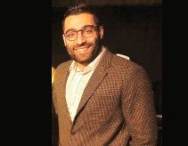
Omid Shearkhani – Studentship – 2015
Omid Shearkhani is a student at the University of Toronto.
About the research
Project title: “Developing and evaluating the performance of an automated computer-aided detection software for the follow-up of metastatic brain tumours”
Project Description
Brain tumours are developed in about 32 out of 100’000 individuals. Out of this number, 75% originated from a region of the body other than the brain, but was spread to the brain (referred to as metastatic brain tumours; MBTs). Furthermore, 20-40% of the individuals with cancer develop MBTs at some point, and as cancer patients live longer, this number is increasing. MBTs can result in mortality and devastating morbidities. Follow-up of MBTs and their changes over time, especially of MBTs smaller in size, remains to be a difficult task, associated with an innate risk of error due to user subjectivity and the demanding workload.
Computer-aided detection (CAD) methods have been successfully used to analyze breast and prostate cancers in magnetic resonance imaging (MRI) scans. To date, no study has focused on developing a CAD system to facilitate MBTs’ follow-up. A software capable of attaining that purpose can serve as a powerful tool in treatment planning and monitoring MBTs, because accurate follow-up of MBTs is necessary for chemotherapy, radiation planning, and surgical approach. The purpose of this study is to develop a CAD software in order to improve accuracy and efficiency for the follow-up of MBTs.
About Omid, in his own words…
 It may not be common to see a medical student who is deeply passionate about mathematics and computer science. Some may even find it paradoxical, because the abstract field of mathematics may seem irrelevant to the practical field of medicine. As a medical student, a technology aficionado, and someone who majored in mathematics as an undergraduate student, I have been amazed by how mathematical models and computer programs are utilized in the fight to conquer brain cancer. It is for this reason that I believe brain tumour research is the field that encompasses all of my interests. It is my ultimate niche, and where my passions and skills intersect.
Being awarded a Brain Tumour Research Studentship, made possible by the generous contribution from kind donors, means that I can pursue a research project in the field that I am truly passionate about, and to utilize my skills and experiences in order to have a positive impact, however small, on the lives of many patients who are battling with brain tumours. This opportunity will allow me to grow as a scientist and future physician, and will thus prepare me for a career as a clinician scientist.
Project Update, May 2016
Initially our technique relied on intensity-based approaches for detecting changing metastatic brain tumours across MR scans. In such approach, after aligning two consecutive scans of the brain of the same patient at different time points, the intensity of respective voxels on each volume is compared. Tumour tissue in an area that previously did not have tumour tissue (i.e. tumour expansion) would result in an increase in intensity on the second scan, and shrinkage of tumour would result in decrease of voxel intensity on the second scan. This approach, however, resulted in a high false positive rate, since other hyperintense structures such as pulsating vessels and movement of dura results in similar changes in intensity (as tumours) across scans. The only approach to minimize such false positives would be to use machine learning techniques which would be resource intensive. To avoid this we looked into the literature for alternative approaches, and were able to find a very suitable approach called deformity-based approach. In this method the deformity filed that results from non-linear co-registration of the two scans is quantified. This quantification gives information about the compression and expansion of the structures that are present in the two scans. After implementing this method, we were able to achieve promising results. Our plan is to focus solely on reducing false positives to improve our current technique so that it has the sensitivity and positive predictive value that is required for use in clinical settings.